How Foundation Models Are Transforming Pathology AI
Foundation models are reshaping how we think about medical data labelling and training. These large, pre-trained models are built to understand general patterns in pathology images and can be fine-tuned for specific diagnostic tasks. Instead of training a new model from scratch for every use case, teams can now start with a high-quality base model that already understands key tissue features.
Large-scale models like UNI, CONCH, and PRISM2 have been trained on hundreds of thousands of WSIs, as well as tissue patches and clinical metadata, to deliver robust performance on dozens of diagnostic and prognostic tasks with minimal task-specific fine-tuning. These models dramatically reduce the need to train from scratch, offering pretrained encodings that understand tissue morphology and context. Instead of investing in model development for each use case, teams can start with a high-quality foundation model already tuned to visual features common across pathology tasks, unlocking faster and more accurate medical data labeling workflows.
The Rise of Multimodal Pathology Intelligence
Even more exciting is the shift toward multimodal learning. This approach combines image data with textual and clinical metadata, allowing models to learn in context. For example, combining whole slide images (WSI) with biomarker reports, pathology notes, and genomic data can lead to far more robust models that mimic the way pathologists interpret complex cases. In practice, multimodal systems can transform medical data labelling by ensuring that annotations are enriched with both visual and contextual information, bringing them closer to real-world diagnostic reasoning.
- Vision-language models
Combine WSIs with textual data, such as pathology reports or clinical notes, to help the model learn both visual patterns and their corresponding clinical meanings. - Vision-knowledge graph models
Link image features to structured medical ontologies or relationships (e.g., disease-tissue associations), enabling the model to reason across hierarchical or semantic clinical data. - Vision-gene expression models
Combine histological images with molecular data such as RNA-seq or biomarker profiles, allowing the model to predict or correlate tissue appearance with underlying genetic activity.
For example, PRISM2 was trained on 2.3 million WSIs paired with real-world diagnostic reports using a two-stage vision-language training strategy. It enables clinical dialogue, diagnostic Q&A, and zero-shot classification, outperforming earlier slide-level models while generalizing across tissue types and diseases.
These multimodal advances begin to mirror how pathologists make decisions: synthesizing visual cues with structured data and medical context.
Case in Focus: PathChat
One emerging initiative in this space is PathChat, a prototype framework being scoped with a major life sciences company. PathChat explores the integration of foundational WSI models with multimodal feedback loops and AI-human interaction. The idea is to enable real-time collaboration between annotators, pathologists, and AI agents.
In practice, this could look like an expert interacting with a digital assistant that references similar cases, highlights key features in the image, and even auto-suggests annotation boundaries. By combining visual understanding with structured clinical context, systems like PathChat move toward a future where annotation is no longer a one-way manual task but a two-way, intelligent exchange.
What It Means for AI Teams
Why this matters for pathology AI builders:
- Build faster with pretrained models that already understand histological patterns and tissue context.
- Train more accurately by combining image, clinical text, and biomarker inputs through multimodal architectures.
- Collaborate better with AI assistants that offer interactive support and smart suggestions to pathologists.
- Scale clinically, with focused annotation needs and models that generalize across labs, protocols, and disease types.
- Support regulatory readiness, attention shifts from solo tools to integrated pipelines with built-in auditability and explainability, particularly for medical data labelling in regulated healthcare environments.
What It Takes to Power These Workflows
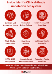
For multimodal and foundation-model-based systems to succeed, the underlying data pipelines need to be incredibly robust. At iMerit, we have built a delivery ecosystem that supports both high complexity and high volume, with a focus on clinical reliability.
Key capabilities include:

- Clinician-Led Annotation Teams with US board-certified pathologists and medically trained annotators.
- Curriculum-Driven Training that enables annotators to develop deep expertise across diverse pathology tasks.
- Smart Tooling with Ango Hub, offering multi-resolution slide viewing, quadrant-based loading, and real-time quality control.
- Multi-Modal Annotation capabilities to combine histological images with biomarker data, pathology notes, and genomic information.
- Dual Shore Delivery for operational flexibility and rapid scaling across regions.
- Multistage Quality Oversight to ensure accuracy and consistency at every stage of the annotation process.
- HIPAA & ISO Compliance to meet stringent privacy and security standards for medical data labelling.
- Domain-Tuned Automation Tools that accelerate workflows without compromising clinical precision.
- Regulatory Ready Workflows to support auditability, traceability, and compliance for research and commercial deployments.
This infrastructure is critical for building ground-truth datasets that meet both research and regulatory requirements. It also provides the flexibility needed to support large, distributed projects where turnaround times and quality expectations are high.
Looking Ahead
As digital pathology continues to evolve, so do the expectations placed on AI models. The future will demand systems that can learn from multiple modalities, adapt to new diseases and staining methods, and deliver results that are both explainable and clinically valid.
This evolution is not just about better algorithms. It is about smarter annotation pipelines, tighter integration between tools and people, and a deep understanding of what clinical quality means in the context of machine learning.
At iMerit, we are committed to supporting this next phase of pathology innovation. Whether it is scaling annotation for a new biomarker, validating foundation models, or enabling human-AI collaboration through initiatives, we combine advanced tooling on Ango Hub, domain-tuned automation techniques, and an expert-led workforce to deliver the domain expertise and delivery maturity needed to help teams move from proof of concept to real-world impact.
Let’s Build the Future of Pathology AI Together
Learn more about our digital pathology solutions here.
Schedule a Demo or Contact Us to explore how we can help you scale digital pathology AI with clinical precision.


